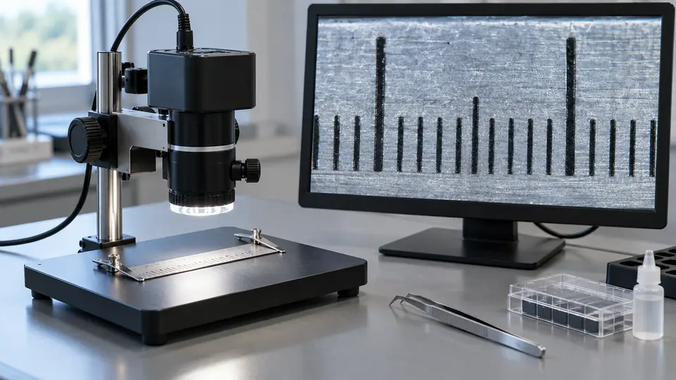
Digital microscope magnification can describe several different things, and those measurements are not interchangeable. A microscope may have optical magnification from its lens system, digital magnification applied by software, or apparent magnification created by displaying an image on a large screen. A high number printed on a product listing does not automatically mean that the microscope resolves more fine detail.
The useful question is not simply, “How many times does it magnify?” It is, “How much genuine specimen detail can the system resolve at the working distance and screen size I will use?” That answer depends on the optics, sensor, resolution, lighting, focus, field of view, and display.
If you are comparing equipment, our guide to digital microscope cameras explains how viewing options, focus, lighting, and camera type affect the choice alongside advertised magnification.
What Magnification Means in a Digital Microscope
Magnification is the enlargement of an object’s image. Resolution is the ability to distinguish two closely spaced details as separate features. Magnification makes an image larger. Resolution determines whether the enlarged image contains additional visible information.
For example, enlarging a captured image with software can make a scratch appear bigger on the screen. It cannot restore surface detail that the camera never resolved. The same principle applies to a microscope that enlarges a live image digitally.
A complete digital microscope system may involve several stages:
- Optical magnification: Enlargement produced by the lens system before the image reaches the sensor.
- Sensor sampling: The camera sensor converts the optical image into pixels.
- Digital magnification: Software crops or enlarges the captured image.
- Display magnification: The image appears larger or smaller depending on the monitor, phone, tablet, or built-in LCD.
These stages can produce very different magnification figures. A specification that says “1,000×” may refer to a manufacturer’s display setup, a digital zoom range, an estimated total magnification, or a measurement convention that is not fully explained. Unless the basis of the figure is documented, it should be treated as a general marketing specification rather than a direct measure of optical detail.
Optical Magnification Versus Digital Magnification
| Type | How It Works | Effect on Detail |
|---|---|---|
| Optical magnification | Lenses enlarge the specimen image before it reaches the sensor or eyepiece. | Can reveal more detail, provided the optical system, illumination, and sensor can resolve it. |
| Digital magnification | Software enlarges, crops, or resamples the captured image. | Increases image size without necessarily adding new specimen detail. |
| Display magnification | The image appears at a particular size on a screen viewed from a particular distance. | Changes apparent size. It does not by itself establish optical resolution. |
Optical Magnification
Optical magnification comes from the microscope’s lens arrangement. A digital stereo microscope may use interchangeable objectives or a zoom optical system. A camera attached to a compound microscope receives an image formed by the microscope objective and may include additional camera-adapter optics.
Optical magnification must be considered alongside numerical aperture, optical quality, sensor sampling, illumination, and specimen contrast. Increasing lens magnification without sufficient resolving power can produce a larger but soft image. Microscopists sometimes describe this as empty magnification, meaning the image is enlarged without a proportional increase in visible detail.
Digital Magnification
Digital magnification enlarges the image after capture or during live video processing. Depending on the software, it may crop the image, interpolate pixels, or resample the frame to fit the display.
Digital zoom can be useful for positioning a small feature, reviewing a captured image, or filling more of the screen. It has practical limits. Once the camera has sampled the available detail, additional enlargement produces larger pixels, softer edges, or visible artifacts rather than new optical information.
Display Magnification
A digital microscope viewed on a 27-inch monitor may make an object appear larger than the same microscope viewed on a small phone screen. Viewing distance also changes apparent size. For this reason, display magnification is not a stable optical specification unless the display size, image scale, and viewing conditions are defined.
A built-in LCD can show a large image that is convenient for inspection, but the apparent size of that image should not be confused with the magnification produced by the microscope’s lens.
Why Advertised Magnification Numbers Can Be Misleading
Low-cost USB and LCD inspection microscopes often advertise very high maximum magnification figures. Those numbers may combine the lens, sensor, software, and display conditions. The product documentation may not state whether the figure is optical, digital, or an estimate based on a particular screen.
A 1,000× label therefore does not establish that the microscope can resolve bacteria, individual cells, fine crystal structures, or microscopic manufacturing defects. It also does not establish a specific field of view or working distance.
Useful questions to ask about a magnification specification include:
- Is the stated value optical, digital, or a combined estimate?
- What object size and field of view are available at that setting?
- What working distance remains at the highest useful setting?
- Is the image captured at native sensor resolution or enlarged by interpolation?
- Does the documentation describe the display size and viewing conditions used for the claim?
- Is the stated range available across the entire image, or does sharpness decline near the edges?
If these details are missing, the magnification number offers limited information by itself. Field of view, image examples with documented scale, optical design, and resolution information usually provide a more useful picture of capability.
Magnification Does Not Equal Resolution
Resolution describes the smallest separation between details that the imaging system can distinguish. In a digital microscope, the final result depends on the lens, numerical aperture, sensor, lighting, contrast, focus, image processing, and specimen characteristics.
Numerical aperture is an optical property related to the cone of light accepted by the objective. It is an important factor in resolving power, particularly in compound microscopy. Magnification alone cannot determine resolution. A high-magnification lens with poor illumination or limited optical quality may show less useful detail than a lower-magnification system with better optics and contrast.
Sensor resolution also has limits. A camera may save a large image file, but the file can contain interpolated pixels or enlarged versions of the same captured information. Native sensor resolution indicates how many photosites are used to sample the image. It does not, by itself, prove that the optics deliver equivalent detail across those pixels.
The same distinction applies to video. A microscope that outputs a 4K signal may produce a 4K-sized video frame, but that does not automatically mean the optical system resolves 4K-equivalent specimen detail.
How Screen Size Changes Apparent Magnification
Screen size changes how large the image appears to the viewer. A small feature displayed across several inches of a monitor may look much larger than the same feature displayed on a phone. The microscope itself has not changed its optical magnification.
Apparent size is also affected by:
- Monitor or LCD dimensions.
- Image scaling settings in the software.
- Full-screen versus windowed viewing.
- Viewing distance.
- Video output resolution.
- Digital cropping and zoom.
This matters for inspection work. A large display can make a solder joint easier to view and can provide more comfortable positioning, but it does not compensate for limited optical resolution. A large, soft image remains soft.
Working Distance, Field of View, And Depth of Field
Magnification affects how much of the specimen appears in the image and how much room remains between the lens and the object. The exact relationship varies by optical design, but higher magnification commonly produces a smaller field of view. The visible area becomes narrower, which can make it harder to inspect a large object or track a moving tool.
Working distance is the space between the front of the microscope’s optical system and the specimen when the image is in focus. It is different from the height of the stand or the distance between the camera and monitor. A short working distance may be acceptable for examining a coin surface, but it can restrict access for tweezers or a soldering iron.
Depth of field is the range of specimen depth that appears acceptably sharp. At higher magnification, the sharp region often becomes thinner, depending on the microscope’s optical design and aperture. A raised component, curved coin surface, or three-dimensional object may therefore require careful focusing or repositioning.
These three properties should be evaluated together:
| Property | What It Describes | Why It Matters |
|---|---|---|
| Field of view | The width and area visible in the image. | Determines how much of a coin, circuit board, or specimen fits on screen. |
| Working distance | The clearance between the lens and the specimen at focus. | Determines room for tools, lighting, and specimen handling. |
| Depth of field | The range of depth that appears acceptably sharp. | Affects curved, uneven, or three-dimensional objects. |
Why Lighting Affects Useful Magnification
Magnification cannot compensate for poor illumination. Fine details may disappear in shadows, glare, low contrast, or excessive exposure. A microscope may enlarge a surface feature while making it harder to recognize if the lighting produces a bright reflection over the same area.
Reflective objects such as coins, polished metal, solder joints, jewelry, and gemstones often require changes in light angle rather than maximum LED brightness. Moving the light, reducing intensity, using diffusion, or changing the specimen angle can reveal surface texture that direct illumination hides.
Biological specimens may require a different approach. Opaque objects are commonly viewed with reflected light, while transparent specimens may require transmitted light and a suitable compound optical system. A USB inspection microscope intended for surface observation should not be treated as a replacement for a compound microscope simply because its advertised magnification is high.
Choosing Useful Magnification For Common Tasks
PCB And Solder Joint Inspection
PCB inspection often benefits from a moderate view that includes surrounding components, stable mounting, adequate working distance, and responsive live video. Excessive digital zoom can make it harder to position a soldering tool or understand the relationship between nearby components.
A high magnification setting may help examine a localized solder joint after the board is inactive and cool. It may be less suitable during active soldering if the lens leaves too little clearance for the tool or if movement becomes difficult to track.
For specific setups that balance viewing area, working distance, and positioning, see our guide to the best digital microscopes for PCB inspection.
Coin Inspection And Photography
A lower setting with a wider field of view is usually more practical for documenting an entire coin. Higher optical magnification can help inspect a localized mark, edge detail, die feature, scratch, or possible surface alteration.
Lighting angle and glare control often affect the usefulness of a coin image more than the maximum magnification figure. A digital microscope can reveal visible surface characteristics, but it cannot establish authenticity, grade, or market value by magnification alone.
To compare options for viewing and documenting coins, explore our guide to the best digital microscopes for coin collecting, including considerations such as lighting and stand design.
Jewelry And Gemstone Observation
Magnification can help show scratches, setting details, surface wear, or visible inclusions. Reflections from polished metal and gemstones may obscure those features, so controlled lighting and specimen positioning are important.
Observation through a digital microscope does not by itself establish gemstone identity, treatment history, authenticity, or value. Those conclusions may require appropriate gemological examination.
Biological Observation
Biological work depends heavily on optical resolution, numerical aperture, illumination, contrast, and specimen preparation. A microscope suitable for observing plant surfaces or insect features may be unsuitable for resolving bacteria or internal cellular structures.
Seeing a small object, resolving its outline, and identifying it are separate tasks. Advertised digital magnification is insufficient evidence for any of them.
How To Evaluate A Magnification Specification
- Identify the microscope type. Determine whether it is a USB inspection microscope, LCD inspection microscope, HDMI system, digital stereo microscope, or camera attached to a compound microscope.
- Separate optical and digital claims. Look for information about objectives, zoom optics, focal range, or software zoom.
- Check the field of view. A useful magnification setting should show an appropriate area of the specimen for the task.
- Check working distance. Confirm that enough clearance remains for tools, lighting, or specimen handling.
- Review native output information. Distinguish sensor resolution, saved image resolution, video resolution, and interpolated output.
- Assess the optical limitation. Look for information about the objective, numerical aperture, focus range, and illumination rather than relying on the largest magnification number.
- Use a scale reference for measurements. Magnification does not replace calibration. Measurements require a reference image captured under the same optical and focus conditions.
Magnification And Measurement Accuracy
A magnified image is not automatically a measured image. Software can display a scale bar or numerical dimension only after the image has been calibrated against a known reference. Calibration may change with zoom, optical configuration, working distance, camera settings, or cropping.
A readout with several decimal places does not prove equivalent measurement accuracy. Display precision and actual accuracy are different. For reliable measurements, use a suitable reference scale, maintain the same microscope configuration, and treat the result according to the limitations of the optical system and calibration method.
Frequently Asked Questions
Is 1,000× Magnification Always Better Than 500×?
No. The higher figure may represent digital enlargement or a display-based estimate. A lower setting with better optical detail, a wider field of view, stronger contrast, and more stable focus may be more useful for inspection.
Does Digital Zoom Reveal More Detail?
Digital zoom can enlarge the captured image, but it does not necessarily reveal additional specimen detail. It may help you view or frame an existing feature. It cannot recover information that the lens and sensor failed to capture.
Can A Digital Microscope’s Screen Size Change Its Magnification?
Screen size changes apparent image size. It does not change the optical magnification of the microscope. A larger display can make an image easier to view, but it cannot create additional optical resolution.
What Is More Important Than Maximum Magnification?
Useful optical detail, field of view, working distance, depth of field, lighting, focus stability, sensor performance, and the intended application are usually more informative than the maximum advertised number.
Can High Magnification Prove That A Microscope Can See Bacteria?
No. Bacterial observation depends on the complete optical system, including resolution, numerical aperture, illumination, contrast, and specimen preparation. Many inexpensive inspection microscopes are intended for surface viewing and are not suitable for detailed bacterial imaging.
The Practical Meaning Of Digital Microscope Magnification
Digital microscope magnification is best understood as image enlargement within a complete optical and imaging system. Optical magnification can project a larger image onto the sensor, while digital and display magnification can make the captured image appear larger afterward. Only genuine resolved detail helps distinguish fine specimen features.
For a useful evaluation, compare magnification with resolution, field of view, working distance, depth of field, lighting, native image output, and the demands of the task. That approach gives a more accurate picture of what the microscope can show than the highest number in its specification list.
